
Stanford-Affiliated Research Brief Revisits 8 Hz “Non-Hertzian Scalar” Exposure In Vitro, Reporting Increased Lymphocyte DNA Synthesis
A research paper attributed to Dr. Glen Rein (Stanford University Medical Center) outlines an in-vitro experiment comparing a “scalar” wristwatch configuration to a standard EM-field control. Using human lymphocytes and DNA-synthesis (thymidine uptake) readouts, the study reports higher proliferation in the scalar condition and calls for clearer dosimetry and replication.
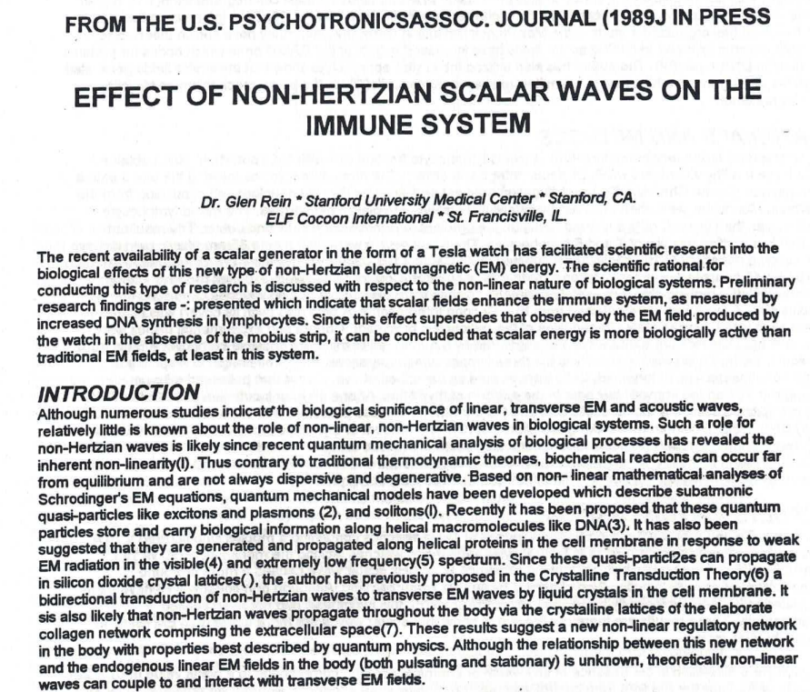
A decades-old lab report is resurfacing because it does something researchers always ask for: it moves an abstract idea into a measurable protocol.
In “Effect of Non-Hertzian Scalar Waves on the Immune System,” the authors describe an in-vitro study design that compares two exposure conditions—one labeled “scalar” and one conventional EM—using a straightforward immune readout: lymphocyte proliferation measured via DNA synthesis.
STUDY BACKGROUND
The paper frames “scalar” energy as a non-linear, non-Hertzian electromagnetic concept and argues that biological systems may respond more strongly to non-linear signaling than to purely linear, transverse EM waves. The research goal is explicit: test whether the proposed field exposure correlates with measurable immune-cell activation in vitro.
HOW THE “SCALAR WATCH” SETUP WAS DESCRIBED
The experiment centers on a commercially described “Tesla watch” approach associated with a Möbius strip configuration. In the paper’s model, the Möbius strip condition is treated as the “scalar” exposure, while a watch configuration without the Möbius strip functions as the EM-field control.
MATERIALS AND METHODS
The protocol described includes:
BLOOD COLLECTION: Whole blood drawn from healthy volunteers.
CELL PREP: Isolation of lymphocytes using standard density-gradient methods.
EXPOSURE: Petri dishes placed under watches inside an incubator, separating experimental and control conditions.
PRIMARY READOUT: DNA synthesis assessed by incorporation of radioactive thymidine into lymphocyte DNA after incubation.
RESULTS: A PROLIFERATION SIGNAL REPORTED
The paper reports that lymphocytes exposed to the “scalar watch” condition showed higher proliferation than those exposed to the control watch condition. It further compares the magnitude of the reported scalar-associated increase to a known chemical immune stimulant (pokeweed mitogen), suggesting the “scalar” condition produced a response in the same general range as chemical stimulation in that in-vitro setup. The table and discussion note statistical significance and variability across trials.
WHY THIS IS RESEARCH-WORTHY (AND WHY IT NEEDS MODERN REPLICATION)
Even for skeptical readers, the most valuable part of this document is not the conclusion—it’s the structure: a defined exposure condition, a defined control, and a defined immune measurement.
At the same time, the paper highlights exactly why this area needs tighter follow-up: field characterization (“dosimetry”), shielding, temperature/humidity controls, device calibration, and blinded handling are crucial if the goal is to distinguish a true effect from experimental drift.
NEXT STEPS FOR A STRONGER STUDY DESIGN
If a modern lab were to replicate this work, a high-quality next phase would typically include:
STANDARDIZED DEVICE CHARACTERIZATION: Independent measurement of emitted fields and environmental artifacts.
SHAM AND BLINDED HANDLING: Randomized assignment and blinded labeling of samples.
DOSE-RESPONSE TESTING: Distance, exposure duration, and signal parameters varied systematically.
PRE-REGISTERED ENDPOINTS: Primary outcomes defined before data collection.
INDEPENDENT MULTI-SITE REPLICATION: Same protocol run by separate teams.
Categories: Sciences, Educational Technology, Healthcare Technology
Tags: DNA, EM-field, Non-Hertzian Scalar Waves, scalar
Additional Links
Just Going Viral
Founded by Hon. Dr. Sandra Rose Michael, DNM, DCSJ, Energy Enhancement System, LLC is a Nevada-based research and technology company dedicated to the responsible exploration of scalar-field and photonic energy phenomena. The company's mission is to p