
Rejuvenations Releases Preliminary Dark-Field Live Blood Microscopy Comparisons Around EESystem™ Exposure, Invites Controlled Replication
Rejuvenations has released preliminary dark-field live blood microscopy images showing pre- and post-session comparisons around EESystem™ exposure (including a 45-minute session and a 2-hour session). The visuals suggest apparent shifts in red-blood-cell shape and aggregation, underscoring the need for standardized protocols and independent, controlled replication.
Rejuvenations is sharing a set of preliminary, case-based microscopy visuals intended to spark a more rigorous conversation: if a field-based environment is claimed to influence biological coherence, what would that look like under a microscope—and how can researchers test it responsibly?
The material presents dark-field live blood microscopy images captured before and after EES/EESystem™ sessions, offering an early, hypothesis-generating look at whether measurable changes may appear in blood-sample morphology over short timeframes.
WHAT WAS DOCUMENTED
The release includes paired image sets labeled as:
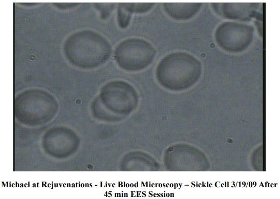
A “before” sample followed by an “after” sample taken after 45 minutes of an EES session (dated 3/19/09).
A separate “before” sample followed by an “after” sample taken after 2 hours of an EESystem™ session (dated 3/25/09).
Across the sets, the “after” images appear to show more uniformly rounded red blood cells and less visible clustering/stacking compared to the corresponding “before” images. These visual differences are presented as observational—not as clinical conclusions.
WHY THIS MATTERS FOR RESEARCH
Live blood imaging is often compelling because it is immediate and visual—but that is also why it demands strong research discipline. Changes in blood appearance can be influenced by many variables, including: sample handling, time on slide, temperature, lighting, hydration status, and interpretation bias.
This is exactly where the opportunity is: these early images can be treated as a starting signal that motivates tighter methodology, clearer metrics, and repeatable testing.
WHAT RIGOROUS FOLLOW-UP COULD LOOK LIKE
To move from interesting visuals to dependable evidence, future work would benefit from:
STANDARDIZED COLLECTION AND TIMING (same finger-stick method, same slide time, fixed imaging window)
CONTROL CONDITIONS (sham exposure, matched duration, randomized order)
BLINDED IMAGE REVIEW (independent reviewers scoring images without knowing “before/after”)
QUANTIFIABLE ENDPOINTS (cell-shape distribution, aggregation scoring, basic hematology markers where appropriate)
REPLICATION ACROSS SITES (different operators, microscopes, and locations)
Categories: Sciences, Educational Technology, Healthcare Technology
Tags: dark-field live blood microscopy, EESystem™, live blood microscopy, red-blood-cell
Additional Links
Just Going Viral
Founded by Hon. Dr. Sandra Rose Michael, DNM, DCSJ, Energy Enhancement System, LLC is a Nevada-based research and technology company dedicated to the responsible exploration of scalar-field and photonic energy phenomena. The company's mission is to p