
Four-Week Hawaii Project Uses Dark-Field Live Blood Microscopy To Explore Pre/Post Patterns During Energy Enhancement System™ (EES) Exposure
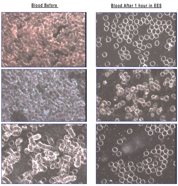
An exploratory November 2004 project in Hawaii captured dark-field live blood microscopy images before and after 1 hour inside the Energy Enhancement System™× (EES). With 36 participants and blood/urine analysis led by Dr. Alvita Soleil, the work spotlights visual patterns that merit standardized, controlled follow-up research.
Curiosity becomes real research the moment you can measure something—consistently, repeatably, and with enough structure that other teams can test the same question.
That’s the spirit behind a four-week live blood cell project funded in Hawaii in November 2004. The initiative brought together 36 male and female participants and used dark-field live blood microscopy to capture visual comparisons of blood samples before exposure and after 1 hour inside the Energy Enhancement System™ (EES). Blood and urine analysis for the project was spearheaded by Dr. Alvita Soleil, Doctor of Oriental Medicine and Director of Kohala Health and Wellness Center.
STUDY OVERVIEW
The project was designed as an exploratory look at whether a structured environment like the EES could correlate with visible shifts in live blood microscopy images. The published materials present paired “before” and “after” image examples intended to prompt deeper investigation—especially around repeatability and measurement standards.
WHAT THE MICROSCOPY IMAGES HIGHLIGHT
The before/after image sets are presented as visual comparisons, where the post-exposure images appear to show differences in cell distribution and clustering patterns versus the pre-exposure images. These observations are not presented as a diagnosis or clinical proof—rather, they function as hypothesis-generating evidence that can help researchers define what to quantify next.
WHY THIS IS RESEARCH-WORTHY
Dark-field live blood microscopy is compelling because it’s immediate and visual—but that’s exactly why research discipline matters. Sample timing, slide preparation, temperature, lighting, hydration, and time-on-slide can all influence what appears in the field of view. If a meaningful signal exists, the fastest way to find it is to tighten the methodology and quantify the outcomes.
WHAT CONTROLLED FOLLOW-UP COULD LOOK LIKE
To move from intriguing visuals to credible findings, a next-phase protocol could include:
Standardized Sample Collection (same method, same timing, same handling)
Fixed Imaging Windows (defined minutes post-collection, defined time-on-slide)
Control Or Sham Comparisons (matched duration and conditions)
Blinded Image Scoring (reviewers unaware of “before” vs “after”)
Quantifiable Metrics (aggregation scoring, shape distribution, repeatability across operators)
Categories: Sciences, Educational Technology, Healthcare Technology
Tags: dark-field live blood microscopy, EES, Energy Enhancement System™
Additional Links
Just Going Viral
Founded by Hon. Dr. Sandra Rose Michael, DNM, DCSJ, Energy Enhancement System, LLC is a Nevada-based research and technology company dedicated to the responsible exploration of scalar-field and photonic energy phenomena. The company's mission is to p